Building a Prototype X-ray Judgment Tool
This guide presents a streamlined approach to creating a prototype X-ray judgment tool using open-source libraries. By utilizing TorchXRayVision alongside Gradio and PyTorch, we simplify the process of analyzing and classifying chest X-ray images. This solution aims to provide users with an interactive experience while ensuring minimal setup requirements.
1. Introduction to the Tool
The purpose of this prototype is to educate users about the functionalities of a medical inference system. It is critical to understand that this tool is not intended to replace professional medical diagnostics.
2. Required Tools
To begin, you will need to install two essential libraries:
- TorchXRayVision: For X-ray analysis.
- Gradio: To create an interactive user interface.
3. Setting Up the Environment
Follow these steps to set up your development environment:
- Install the required libraries using the command:
!pip install torchxrayvision gradio. - Import necessary libraries:
import torch
import torchxrayvision as xrv
import torchvision.transforms as transforms
import gradio as gr
4. Model Initialization
Load a pre-trained DenseNet model, which is essential for the inference process:
model = xrv.models.DenseNet(weights="densenet121-res224-all")
5. Retrieving Pathology Labels
Pathology labels are crucial for interpreting the model’s predictions. If the model does not retrieve these labels, a default list is provided:
try:
    pathology_labels = [model.get_labels()]
except Exception as e:
    pathology_labels = [
        "Atelectasis", "Cardiomegaly", "Consolidation", "Edema",
        "Emphysema", "Fibrosis", "Hernia", "Infiltration", "Mass",
        "Nodule", "Pleural Effusion", "Pneumonia", "Pneumothorax", "No Finding"
    ]
6. Classifying X-ray Images
The classification function processes the X-ray image and returns diagnostic information:
def classify_xray(image):
    try:
        transform = transforms.Compose([
            transforms.Resize((224, 224)),
            transforms.Grayscale(num_output_channels=1),
            transforms.ToTensor()
        ])
        input_tensor = transform(image).unsqueeze(0)  # Add batch dimension
        preds = model(input_tensor)
        pathology_scores = preds[0].detach().numpy()
        results = {label: float(score) for label, score in zip(pathology_labels, pathology_scores)}
        sorted_results = sorted(results.items(), key=lambda x: x[1], reverse=True)
        top_label, top_score = sorted_results[0]
        return f"Prediction: {top_label} (score: {top_score:.2f})nFull Scores:n{results}"
    except Exception as e:
        return f"Error during inference: {str(e)}"
7. User Interface Creation
Using Gradio, create an interface that allows users to upload their X-ray images:
iface = gr.Interface(
fn=classify_xray,
inputs=gr.Image(type="pil"),
outputs="text",
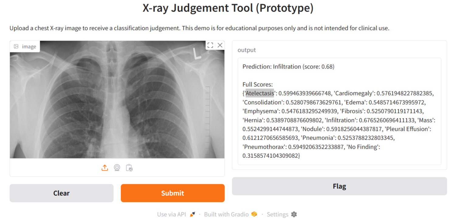
title="X-ray Judgment Tool (Prototype)",
description="Upload a chest X-ray image to receive a classification judgment. This demo is for educational purposes only and is not intended for clinical use."
)
iface.launch()
8. Conclusion
Through this tutorial, we have explored the development of an interactive tool for X-ray analysis. While this prototype is not ready for clinical deployment, it serves as an important foundation for future innovations in medical imaging applications. As you advance, remember to validate your model thoroughly and adhere to medical standards. This prototype offers a clear starting point for enhancing diagnostic practices with artificial intelligence.
For further guidance on integrating AI into your business operations, please feel free to contact us at hello@itinai.ru or connect with us on Telegram, X, and LinkedIn.